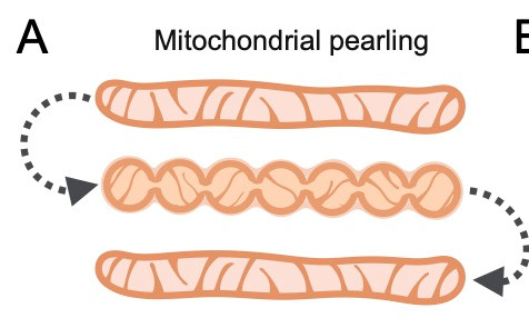
This phenomenon, known as mitochondrial pearling, entails a transient shape change that persists for a short time, sometimes just a few seconds, before reverting back to the normal tubular shape.
Comments
Log in with your Bluesky account to leave a comment
The transient nature of pearling makes it hard to detect - in normal cells you might need to sit and watch one spot for a long time and then, if you blink, you'll miss it
Pearling has been noted before, often in specialized types and especially cases of damaged or dying cells, where the pearling persists over longer time periods. But one thing Gav has found is that pearling is EVERYWHERE
During an internship at Calico, Gav got to work with Kayley Hake and Andy York and their teams to image mitochondria with Snouty. This was an amazing opportunity for learning advanced microscopy, like doing an internship at the Vatican to learn catholicism.
He found that pearling was ubiquitous, occurring in every cell type that he has looked at, including: U2OS, Hek293, COS7, RPE1, Jurkat, primary human fibroblasts primary CD8 T-cells, iPSC derived neurons, and budding yeast (shown here).
Evidently, spontaneous pearling is a universal feature of mitochondrial dynamics, just like fission and fusion, raising (at least) two questions: how does it happen, and what (if anything) is it for?

Comments
https://andrewgyork.github.io/high_na_single_objective_lightsheet/