
🧠 Thread 1: Brain Blood Supply – The Anatomy Behind the Magic!
The brain may run the show, but without blood, it's lights out! 🧠💉 Let's explore the vascular anatomy of the brain—because knowing these pathways can help you localise strokes in seconds. 🧵
#Medsky #MedEd #MedSchool #MedStudent
The brain may run the show, but without blood, it's lights out! 🧠💉 Let's explore the vascular anatomy of the brain—because knowing these pathways can help you localise strokes in seconds. 🧵
#Medsky #MedEd #MedSchool #MedStudent
Comments
🧠 Internal Carotid Arteries (ICA) → ACA & MCA.
ACA: Medial surface (legs) + anterior corpus callosum.
MCA: Lateral surface (face/arms) + language areas.
💡 Lenticulostriate arteries → basal ganglia (stroke hotspot).
🧠 Vertebral Arteries → Basilar artery → PCA.
PCA: Occipital lobe (vision), inferior temporal lobe (memory), & thalamus.
💡 PCA strokes = visual field loss; often subtle initially!
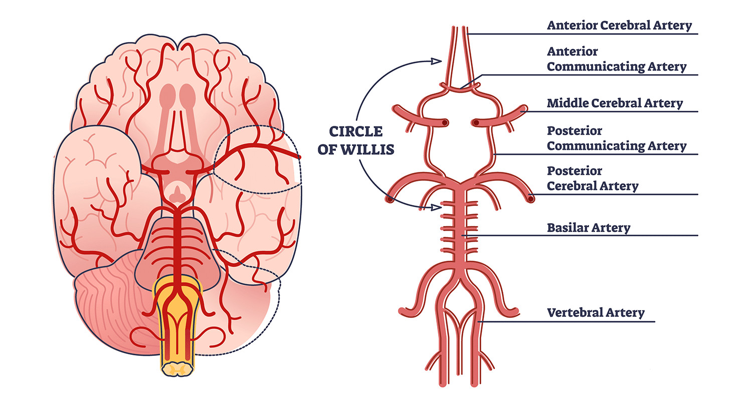
🎯 Circle of Willis: The brain's backup plan!
Anterior communicating artery → joins ACAs.
Posterior communicating arteries → link ICAs & PCAs.
💡 Incomplete in ~50% of people → increased stroke risk.
second image shows common aneurysm sites
💧 Venous Drainage:
Superficial veins → dural sinuses (e.g. superior sagittal).
Deep veins → Great vein of Galen → straight sinus → jugular veins.
🧠 Glymphatic system clears waste—especially during sleep!
❓ Quiz:
A patient has a stroke affecting the medial frontal/parietal lobes, causing right leg weakness.
Which artery is blocked?
A) MCA
B) ACA
C) PCA
D) Basilar
💡 Drop your answers below!
🧠 Real-World Relevance:
In stroke emergencies, knowing vascular territories helps predict symptoms before imaging.
👉 Part 2 tomorrow: Clinical correlations & stroke syndromes!