Profile avatar
nat-prunet.bsky.social
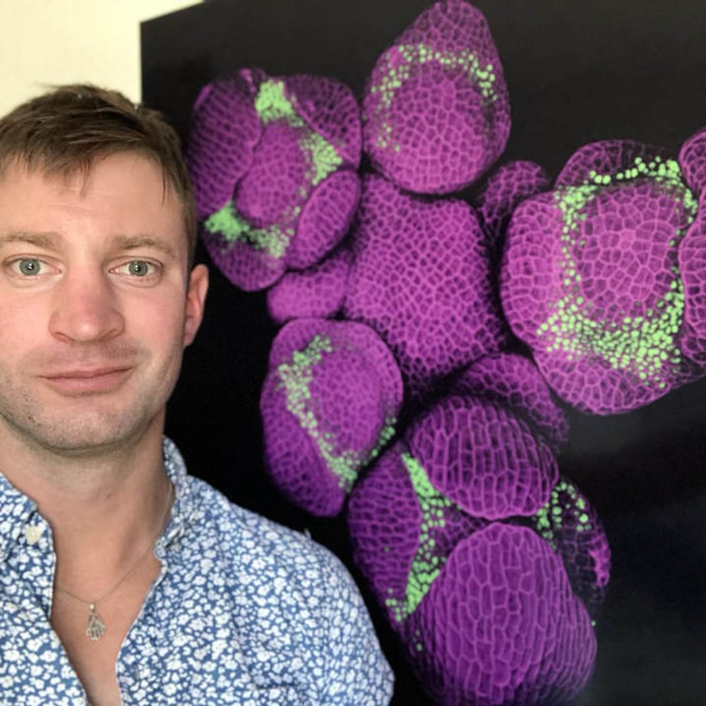
Microscopic farmer 🔬🌺 Microscopy core director @UNC Chapel Hill Former plant developmental biologist
100 posts 657 followers 694 following
Prolific Poster
Conversation Starter

Rhizaria and the Blue Hole 11/2023 22 X 32 inchers [55.9 X 81.3 cm] Colored Pencil on Paper #sciart #gawoski #plankton

Heterokont is the Anagram of Tenterhook 9/2018 13 X 20 inches [33 X 50.8 cm] Graphite on Paper #sciart #gawoski #plankton

Head scales of the Sulcata tortoise, imaged with light sheet microscopy. Our new study in @cp-iscience.bsky.social reveals that their emergence is controlled by both chemical cues and mechanical forces. Read the full article here 🐢🔬🧪 www.cell.com/iscience/ful...

Liver immunostaining for this #FluorescenceFriday

This image highlights the research focus of both my PhD (mitochondria in green) and postdoc (vacuoles in magenta). I love working with plant organelles, they are so dynamic and cool. #plantcell #microscopymonday

Beautiful microscopy of fungi!

This #microscopymonday image is of a #Drosophila male reproductive system: paired coiled testes, seminal vesicles & accessory glands, and a single ejaculatory duct. Each coiled testis is about 1.5mm. Image captured by MDI Bio Lab Sr. Scientist Travis Carney. #standwithscience #researchmatters 🧪 🤝

FINAL CALL – This Thursday, 6pm, in-person 🌿 Step into a hidden world where art meets science under the microscope! Join us for this in-conversation as celebrated visual artist Rob Kesseler FLS reveals how cutting-edge microscopy unlocks the astonishing architecture of plants. bit.ly/4manReO

Tobacco Pair in Red 8/2018 20 X 24 inches [50.8 X 61 cm] Colored Pencil on Paper #sciart #gawoski #pollen

This #confocal image of a #jellyfish by Sosuke Fujita is exquisite! #microscopy #bioart #sciart

😍

Just stumbled upon these #birds I painted when I was 10 back home in France! #birding #birdwatching #painting #bioart #sciart

A perfectly round liver organoid stained for mitochondria (yellowish/green), e-cadherin in blue and nuclei in pink for #MicroscopyMonday

🧠 Missing link of synchronized skull and brain development and evolution revealed — explaining rare congenital syndromes. 🧬 Evolution meets development and medicine. #Evolution #Development #BrainSkull #EvolutionaryInnovations www.evolbio.mpg.de/3823787/news...

Gorgeous!

Artificial feeders have allowed the Anna’s hummingbird to expand their range out of Southern California up to the state’s northern end. They have also driven a transformation of the birds themselves.

After years studying #plant #devbio, I started a #chicken #development experiment 🐣🐥

These are stunning!

😍

😍

Sponge choanoderm belongs in a museum #FluorescenceFriday

Happy #FluorescenceFriday!

😍

Why wait for #FluorescenceFriday if you can have a neat #zebrafish right now. (pic credit Amanda Garfield @cudevbio.bsky.social) #devbio

Love these!

Awesome dehiscence video!

Gorgeous images!

If you need to attend a microscopy or bioimage analysis training course to support your #cellbio research but don't have funding, check out our JCS–FocalPlane Training Grants. 🔬🖥️https://www.biologists.com/grants/jcs-focalplane-training-grants/

😍

One of the reasons I love #DevelopmentalBiology is that it's so visually engaging. I had the pleasure of organising an image competition at the GRC last March, and share this passion. You can now check out these images on @the-node.bsky.social: thenode.biologists.com/grs-developm...

stunning!

🔬✨ Today for #FluorescenceFriday: A close-up of the dorsal part of the first gill arch in a mature zebrafish. The intricate vascular network supporting respiration, visualized using kdrl:mcherry. #Zebrafish #DevBio #Microscopy #Fluorescence #SciArt

Preprint: IntAct-U-ExM enables robust, isoform-specific expansion microscopy of actin networks in yeast and mammalian cells. A cool study led by @anubhavdhar.bsky.social in collaboration with Sudarshan and Deepak Nair. www.biorxiv.org/content/10.1...

Assembling rail microscopes from @thorlabs.bsky.social for my #microscopy course @uncchapelhill.bsky.social. These are super fun teaching tools! #microscope #optics #bioimaging

Another academic year is past and this weekend we said goodbye to the latest set of graduates from UNC Biology. Teaching here is a privilege and seeing so many students excel & getting to know many of them is a yearly highlight. Biology holds its own ceremony, allowing us to honor them by name 1/n

A Dark sectioning algorithm removes background and provides single-frame optical sectioning in fluorescence microscopy, facilitating improved deep-tissue segmentation accuracy. www.nature.com/articles/s41...

😍

Simultaneous imaging of twelve #zebrafish embryos using Light Sheet Fluorescence Microscopy (LSFM). Credit to Prof. Jan Huisken & Flamingo Microscope (huiskenlab.com/flamingo/). #ZebrafishZunday 🧪

The Analytical and Quantitative Light Microscopy (AQLM) course has come to a close. For two weeks, researchers examined the theory of image formation and the subtle interactions between light and a specimen. Learn more about this and other courses here! bit.ly/3GMpPSn

🧫The #heterogeneity of gastrointestinal (GI) #organoids: Image of the Month📆 🔬Patient-derived #cancer & healthy #organoids imaged using spinning disk confocal #microscopy to link 3D morphology to outcomes. 🎨 By Sanika Jahagirdar @sanikajah.bsky.social from @tudresden.bsky.social #sciart #bioart