cellarchlab.com
cellarchlab.com Univ. of Basel Biozentrum🇨🇭. Exploring molecular architecture inside cells with #CryoEM #TeamTomo. Plants and algae in a changing climate. ❄🔬 OF 🌿 4THE 🌍!
697 posts
4,880 followers
2,210 following
Regular Contributor
Active Commenter
comment in response to
post
The crowded cellular environment of physcomitrella….
comment in response to
post
algae rule! all this sex determination stuff would also be interesting to our new colleague Claudia Keller Valsecchi
comment in response to
post
Congrats Jim and crew! 🙌
comment in response to
post
Congrats Sven! You are going to do amazing things in the world of #TeamTomo and beyond 🤗
comment in response to
post
I can smell this photo. Wild sage after the rain.
comment in response to
post
COOL! I remember seeing so many empty shells of these when sampling with @embl.org TREC. But I never found a living one...
comment in response to
post
Haha totally. Dose symmetric tilt scheme. #TeamTomo
comment in response to
post
OoooOOOooo 😍
comment in response to
post
Yeah cool! But is it surprising? If a 200 kDa structure like a 10 nm nucleosome has so many pixels and details for a high-res average, couldn’t a 25 kDa 1-2 nm structure be a dot? That’s a couple pixels at bin4. Such dots are everywhere in cellular tomos. We can’t assign protein IDs to dots though🔎⚫️
comment in response to
post
Yes totally! 💯 👍
comment in response to
post
In many of the salty algae we’ve looked at, the concentration of discrete densities isn’t higher. But their contrast against the cellular background is lower. Filamentous cyanobacteria have better contrast than single celled cyanobacteria. The cell buffer mysteries continue… 🕵️♂️🔎
comment in response to
post
Sorry if that wasn’t clear. So in high-quality cellular tomos, density is localized to discrete dots for all structured objects >20 kD. The different “cell juice” contrast must come from smaller stuff between. Disordered proteins could contribute, but I doubt that varies dramatically between species
comment in response to
post
Hi Reika😀 They're monomers. Actually the VIPP1 is tagged with mCherry (monomeric, 28 kDa). I mean, the nucleosome (200 kDa) is chunky enough to enable a sub-nm subtomo average. So 25 kDa proteins being dots should not be surprising. Seeing them well of course depends on the "cell buffer contrast"
comment in response to
post
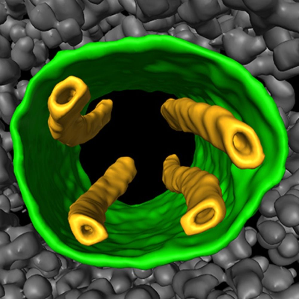
Nice question! Actually, yes a 27 kDa protein (GFP) is visible as a distinct dot in tomograms (even with an old K2 detector). See all the dots decorating PolyQ filaments (green arrowheads, Bäuerlein et al.) and VIPP1-coated membrane tubes (panels O-R, Gupta et al.). The "cell buffer" is smaller! 🧪🧶🧬
comment in response to
post
And modern tomograms such as the Chlamy dataset are much more crisp that those in the above papers (Falcon4i detector, pixel size of 2Å instead of 3.5Å, -1 to -3 um defocus instead of -5 to -8). The details can make your eyes bleed if you stare too long at current tomos 👁️👁️😅
bsky.app/profile/cell...
comment in response to
post
Here's the papers. I'm sure there are more examples of visualizing small known denities in cellular tomograms #cryoET 😁
@felixbauerlein.bsky.social et al., 2017
pubmed.ncbi.nlm.nih.gov/28890085/
Gupta et al., 2021 (with @svenklumpe.bsky.social @wojwie.bsky.social)
pubmed.ncbi.nlm.nih.gov/34166613/
comment in response to
post
Nice question! Actually, yes a 27 kDa protein (GFP) is visible as a distinct dot in tomograms (even with an old K2 detector). See all the dots decorating PolyQ filaments (green arrowheads, Bäuerlein et al.) and VIPP1-coated membrane tubes (panels O-R, Gupta et al.). The "cell buffer" is smaller! 🧪🧶🧬
comment in response to
post
I also want to point out that while Chlamy is the poster child for light “cell buffer” and high contrast, it’s obviously not the only cell with these properties. It’s an interesting phenomenon and something to consider when picking cells or conditions (if your question is flexible and allows choice)
comment in response to
post
I think exactly this!☝️ So far, the salty bugs we’ve looked at are not as contrastic as Chlamy. And the extreme— I remember how horrible the contrast was in a halophile @pilhoferlab.bsky.social was studying. Other types of cells (yeast etc.) may have dark “cell buffer” for other reasons. Interesting!
comment in response to
post
Number of coding genes does not equal copy number. Yeast does have a higher density of ribosomes in the cytosol (2x higher?). But every cell I’ve ever looked at is crowded with complexes. The issue is variety since only ~100 complexes or so are abundant enough for easy averaging. Many are rare🔎
comment in response to
post
@wallaceucsf.bsky.social always used to call it that 💚. But of course, I think it's so much more. It also swims! And eats light! And has an eye! Understanding the bioenergetics relationships between mito and chloro are certainly interesting though @florentwaltz.bsky.social
comment in response to
post
woooooo!! 👩🔬👨🔬
comment in response to
post
IMHO the contrast in human cells falls somewhere in the middle, but it's cell-type dependent. Neurons have nice contrast (maybe makes sense since they aren't still growing). But RBCs🩸 are the worst (despite low complexity) because of all the iron-- ask Leonie & Mimi www.biorxiv.org/content/10.1...
comment in response to
post
Many "simple" bacteria also have poor contrast. It's not about complexity or variety of complexes. I'm not an SPA guy, but I've taken micrographs of proteins without washing away sugar from a gradient- bad contrast! All heavy atoms scatter electrons, be it proteins, sugars, lipids, salts, whatevs...
comment in response to
post
To compare Chlamy with yeast...
cell diameter: ~10 vs. ~4 um
coded proteins: ~15k vs. ~5k
contrast: superb vs. dark
Yeast are packed with ribosomes, but they also have a high turgor pressure and I would guess a high concentration of electron-dense solute (in this case maybe sugars & amino acids)
comment in response to
post
Chlamy lives in a low osmolarity environment (fresh water) and must constantly pump out large volumes of water through it's contractile vacuoles, otherwise it will explode. To quote @sofie-dot-rec.bsky.social, it's a water ballon🎈. But that's just the "buffer" between the complexes.🧪🧶🧬
comment in response to
post
Chlamydomonas is easy and grows fine in many conditions. Also in ambient light or darkness (provided with a carbon source). Agree it’s not a model for everyone (is there a universal one?) but it’s not bad :). Here’s a nice intro for new fans of Chlamy: pubmed.ncbi.nlm.nih.gov/31189738/
comment in response to
post
You got it! We have such a backlog of mysterious structures we are curious about. I don't think Chlamy has a much lower density of complexes. My current theory is just that the "buffer" of the cell is more watery. Like, you wouldn't want to do SPA with too much sucrose or salt still in the buffer...
comment in response to
post
The most beautiful apoferritin 😅🥰
bsky.app/profile/cell...
comment in response to
post
I understand that we use averages to not freak out about specific yearly fluctuations, but do you feel a 10-year average is still the best metric? It lags behind quite a bit. Would a 5-year average be more reflective of the current rate of warming (while still resistant to a 1 or 2 year bump)?
comment in response to
post
Beautiful! Congrats!